High-Content Screening: Designing Imaging Assays for Phenotypic Hit Selection
High-content screening is useful when a compound's effect cannot be understood from one averaged signal alone. Imaging can reveal where a response occurs, which cells respond, and whether an apparent benefit accompanies cell loss or abnormal morphology. This guide helps discovery teams decide when to commission an imaging assay, define an analysis package, and select phenotypic hits that justify confirmation rather than advancing compounds on attractive images alone.
01 Scope and Rationale
When does high-content screening add decision value?
Consider high-content screening when location, cell state, or population heterogeneity changes the meaning of the result. Examples include nuclear translocation, redistribution of a cellular marker, changes in organelle morphology, or a response restricted to a subpopulation. If the relevant distinction can be measured reliably with a simpler endpoint, additional imaging may not improve the decision. Begin with the information needed to distinguish useful compounds from misleading responses.
A plate reader usually summarizes a signal across a well. Image analysis can preserve measurements at the level of fields, individual cells, or cellular compartments before those measurements are summarized. That extra resolution is valuable only when acquisition and analysis are appropriate. More channels and more features also create more opportunities for measurement error, confounding, and unstable interpretation. The project should specify which information will influence compound selection.
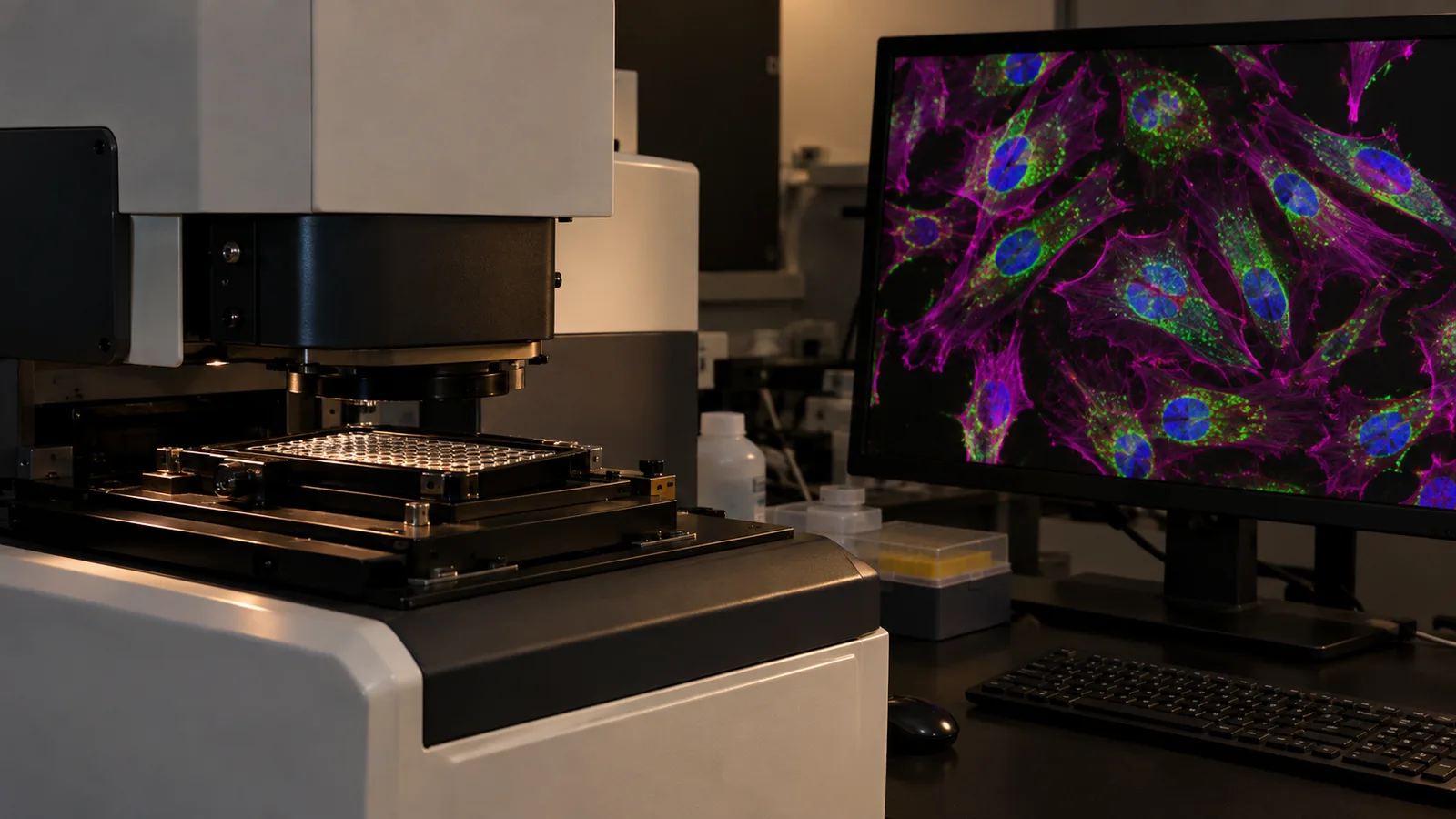
Creative Biostructure's high-content screening services combine automated fluorescence imaging with image processing and data analysis. A suitable starting brief describes the cellular response, existing assay evidence, and the ambiguity that imaging should resolve. The initial discussion can then address whether a focused endpoint assay or a broader phenotypic profile is appropriate for the question and available model.
Focused imaging assays and morphological profiling serve different purposes
A focused imaging assay measures a predefined event, such as a change in nuclear-to-cytoplasmic marker distribution. Its primary endpoint should have a clear biological interpretation and a practical route to confirmation. Broader morphological profiling collects multiple characteristics to describe cellular responses and compare compounds. Such profiles can generate useful similarities and differences, but they do not identify a direct molecular target by themselves.
Cell Painting is a published example of multiplexed morphological profiling. It illustrates how coordinated staining and image analysis can describe several cellular components. It should not be treated as a synonym for every HCS experiment or as an automatically included service configuration. A project requesting a particular staining protocol, cell model, or analytical pipeline needs a feasibility review and an agreed scope before those elements become commitments.
Machine learning can help analyze rich image data, but it cannot repair a comparison that confounds treatment with batch, plate position, or acquisition conditions. Ask what the model is intended to predict and how that prediction will be checked on independent experimental material. A useful service output explains the evidence behind compound prioritization; the presence of an AI component is not an endpoint or a substitute for biological controls.
Define a phenotype that can survive retesting
Write the desired response as an observable pattern with boundaries. For example, a hypothetical project might seek increased nuclear localization of a marker without a substantial decline in intact cell number. This is more informative than requesting compounds that increase fluorescence. The latter could select brighter staining, altered cell size, or accumulation of damaged cells rather than the intended biological event.
Specify whether the decision depends on an average shift, the fraction of responding cells, or a rare phenotype. Those objectives require different sampling and analysis choices. A well average can conceal a meaningful minority response, while a few unusual cells can distort an unstable metric. The assay should collect enough relevant observations to support the chosen endpoint without assuming that every cell in a well is an independent experimental replicate.
| Project question | Useful imaging output | Important alternative explanation |
|---|---|---|
| Does a marker move between compartments? | Validated compartment masks and a location-sensitive measurement. | Segmentation errors or changes in cell shape can alter apparent localization. |
| Does treatment restore a disease-associated morphology? | A predefined comparison with disease and reference conditions. | General stress or selective loss of abnormal cells can resemble rescue. |
| Do only some cells respond? | Response distributions and a justified responder definition. | Sampling or uneven staining can create apparent subpopulations. |
| Do compounds produce similar phenotypic profiles? | Reproducible profiles compared under matched experimental conditions. | Shared toxicity or batch effects can produce similarity without a shared target. |
| Which hits merit confirmation? | Concentration-dependent phenotype with image QC and cell-health context. | A strong image-derived score may disappear when acquisition or analysis changes. |
02 Build an Imaging Screen Around a Reproducible Phenotypic Decision
The acquisition and analysis plans should be developed together. Define what will be measured, how unreliable images will be identified, and how a candidate response will be challenged before committing to a large image collection.
-
1
Specify the cell model, perturbation, and primary endpoint
Describe the cell system and why it can display the relevant biology. Record the treatment context, exposure period, marker strategy, and expected direction of change. A model that is convenient to image may not reproduce the pathway or state needed for the project. Identify which model limitations are acceptable for initial screening and which would undermine the intended conclusion.
Separate the primary endpoint from supporting measurements. Cell count, nuclear morphology, and intensity distributions may help interpret the principal response without becoming interchangeable definitions of success. When the underlying measurement is not established, assay development and optimization can be scoped before screening. This prevents a large campaign from becoming an expensive search for an endpoint after the images have already been collected.
-
2
Establish acquisition conditions that preserve the relevant information
Choose channels, magnification, field coverage, and exposure settings around the biological distinction. Too few fields may miss a heterogeneous response; excessive acquisition can increase storage and analysis demands without changing the decision. Review representative images from controls and expected response conditions. Confirm that the signal is measurable without routinely saturating pixels or losing the structures required for analysis.
For fixed-cell experiments, assess preparation and staining consistency. For live-cell imaging, consider whether the observation schedule and imaging conditions affect the cells. The exact configuration should follow feasibility findings rather than a universal prescription. Record instrument settings and sample preparation details so later comparisons can distinguish biological changes from differences introduced during imaging.
-
3
Validate segmentation before trusting derived measurements
Segmentation defines the objects being measured, such as nuclei, cells, or intracellular regions. Inspect masks overlaid on images from several relevant conditions, including strongly affected cells. A pipeline that works on untreated cells may fail when treatment changes shape, density, or texture. Incorrect boundaries can create an apparent concentration response even when the intended biological marker has not changed as assumed.
Agree on how merged objects, fragments, out-of-focus fields, and debris will be handled. Exclusion rules should be visible and applied consistently. Do not remove unusual cells simply because they look different from the controls; they may represent the phenotype of interest. Distinguish an analysis failure from a genuine morphological response and retain the evidence needed to review that distinction.
-
4
Design plates and replicates to separate treatment from nuisance variation
Distribute controls and test conditions so that position or batch does not become a substitute for treatment identity. Record plate, well, acquisition order, and relevant preparation information. A compound tested only on one unusual plate can appear distinctive for reasons unrelated to its pharmacology. The experimental design should make the main comparison identifiable rather than relying on software to remove every nuisance factor afterward.
Plan biological and technical replication according to the intended conclusion. Thousands of cells from one well do not supply thousands of independent treatment experiments. Summaries and uncertainty should reflect the experimental unit. For profiles, test whether similarities persist across independent runs instead of judging them only from a visually separated cluster in a single projection.
-
5
Pilot the analysis and freeze the main selection rules
Use a bounded pilot to evaluate image quality, endpoint behavior, cell-health measures, and analysis stability. Examine how sensible changes to thresholds or segmentation affect the ranking. If a candidate is selected only under one fragile setting, that sensitivity should be reported. Establish the main hit definition and review process before applying them to the larger collection.
Match the collection to the objective through compound library design and preparation when selection is still open. A mechanistically annotated set can support reference comparisons, while a diversity-oriented set addresses a different discovery question. Library composition changes what a phenotypic result can teach you and should be considered alongside imaging capacity and follow-up resources.
-
6
Retest phenotypes with concentration and cell-health context
Confirm shortlisted responses using an agreed concentration series and suitable repeat material. Review the phenotype alongside cell number, relevant health indicators, and the underlying images. A response observed only near widespread cellular disruption deserves a different interpretation from a reproducible effect within a usable experimental range. Concentration dependence helps characterize a phenotype but does not alone establish specificity.
Select a complementary experiment that tests the most plausible alternative explanation. Depending on the question, this might be another marker, a different detection method, or a functional readout. The purpose is to challenge the proposed interpretation, not to accumulate similar images. Keep confirmed responses, ambiguous responses, and technical failures distinct in the final shortlist.
-
7
Connect phenotypic evidence to the next biological question
Decide whether the next step is model confirmation, chemical follow-up, pathway analysis, or target investigation. A robust image phenotype may justify further work even when its molecular mechanism remains unknown. Conversely, similarity to a reference compound should not be reported as proof that both compounds bind the same protein. Describe what the imaging establishes and what remains a hypothesis.
For an active compound whose target is unresolved, reverse docking and target fishing can support a separate hypothesis-generation stage. Computational target proposals should be evaluated with independent experimental evidence. The imaging dataset supplies biological context for that stage; it does not convert a predicted interaction into a validated mechanism.
03 Common Pitfalls and How to Avoid Them
-
Calling cell loss a therapeutic rescue
A treatment can reduce an undesirable signal by eliminating the cells that produce it. Review intact cell counts and relevant health measurements alongside the desired endpoint. Where possible, inspect whether surviving cells display the intended change rather than relying on a lower total well signal. A putative rescue that depends on substantial loss of the measured population requires explicit qualification and an appropriate follow-up experiment.
-
Equating feature quantity with biological insight
A long feature table can contain redundant, unstable, or poorly interpretable measurements. Prioritize features that are reproducible and relevant to the comparison, and document how they are summarized. Adding measurements after inspecting which ones favor a preferred compound can bias selection. Exploratory findings can still be useful, but they should be labeled and tested separately from the predefined screening endpoint.
-
Treating a clustering plot as mechanism confirmation
Nearby points in an embedding indicate similarity under a particular representation and analysis, not necessarily shared pharmacology. Changes in normalization, feature selection, or cell health can alter the apparent groups. Review the underlying measurements, reference compounds, and reproducibility. A plot is a useful navigation tool for a dataset; it is not an independent experiment establishing a molecular target.
-
Losing the connection between images and numerical results
A spreadsheet of well scores is difficult to audit if the corresponding images, masks, and processing settings cannot be located. Preserve identifiers across acquisition and analysis. Agree on which raw and processed files will be supplied, retained, or made available for review. Representative images should explain the analysis and illustrate typical observations rather than showing only the most visually convincing fields.
An HCS shortlist should state why each compound remains interesting after image quality and cell-health review. A strong phenotype with an unresolved mechanism and a weak phenotype with a familiar annotation are different situations; neither should be promoted automatically by a single composite score.
04 Deliverables
Define the data handoff before acquisition. Imaging projects can generate substantially more material than a conventional endpoint assay, so file access, identifiers, analytical transparency, and interpretation should be part of the agreed scope.
-
Acquisition record and image-quality assessment
Request the cell preparation, treatment conditions, plate maps, acquisition settings, and relevant QC summaries. Specify how excluded fields or wells are documented. The record should allow a reviewer to understand whether the dataset consistently captures the intended structures and how technical failures affect the comparisons. Agree on image delivery and retention rather than assuming that every raw file is included automatically.
-
Analysis definition and representative segmentation checks
Document the measured objects, feature definitions, processing sequence, and important parameter choices. Include representative overlays from controls and affected samples. For model-assisted analysis, describe the model's role and the validation relevant to the project. The deliverable should make the relationship between image content and reported endpoint understandable without requiring the client to infer it from software output alone.
-
Compound-level results with supporting distributions
Provide traceable compound, concentration, replicate, and well identifiers together with the agreed summaries. Where heterogeneity matters, include distributions or responder fractions rather than only a mean. Retain flags for low cell count, image defects, and other interpretation limits. A reviewer should be able to distinguish a consistent moderate response from an unstable value driven by a few poorly measured objects.
-
Prioritized phenotype report and confirmation options
State which compounds meet the predefined criteria, which require clarification, and which fail technical or biological review. Explain the evidence supporting each category and propose the next discriminating experiment. This is more useful than a ranked list without reasons. Any suggested mechanism should be labeled according to its evidence, particularly when it is inferred from phenotype similarity rather than measured directly.
A useful outcome may be a shortlist, a revised endpoint, or a finding that the current cell model cannot answer the question reliably. Judge the project by the quality of the decision it enables. A large image archive or a high number of apparent hits does not establish that the screen has produced interpretable starting points for discovery.
05 Planning a High-Content Screening Project with Creative Biostructure
Bring the biological ambiguity into the initial discussion
Creative Biostructure supports automated imaging and analysis for cellular readouts including protein translocation, viability, apoptosis, cell-cycle changes, and related responses. The appropriate configuration depends on the model and endpoint. Share the existing evidence and explain which interpretation is currently uncertain. A request to distinguish localization from expression, for example, leads to a different design from a request to compare broad morphological responses.
Provide representative images where available, along with cell identity, treatment conditions, marker information, and known references. Unsuccessful images are also useful because they reveal density, staining, or segmentation problems. If the project lacks an established assay, scope a feasibility stage with a concrete question and decision point. Avoid treating a discovery pilot as if it were already a fully qualified production screen.
Compare proposals by interpretable coverage and follow-up value
The main cost drivers can include assay development, acquisition settings, channels, fields, concentration coverage, replication, image storage, and analytical complexity. A lower per-well price may omit the information needed to distinguish toxicity from the desired response. Compare proposals using the same biological endpoint and data handoff requirements rather than comparing image counts alone.
Agree on who reviews ambiguous phenotypes and how additional analysis or retesting is authorized within the project scope. Separate the initial screen from downstream mechanism studies. This makes it possible to commission the imaging work at an appropriate scale while retaining a clear path to deeper characterization when the evidence supports it.
06 Frequently Asked Questions
Is high-content screening always better than a plate-reader assay?
No. It adds value when spatial information, morphology, or cellular heterogeneity affects the decision. A simpler assay may be preferable when a reliable aggregate endpoint answers the question. Compare the biological information gained with the additional acquisition, analysis, and confirmation requirements before choosing the format.
Can HCS distinguish a useful response from cytotoxicity?
It can help when the assay includes appropriate cell-health context and interpretable images. It does not automatically remove that ambiguity. Review the desired endpoint alongside cell count, morphology, and relevant supporting measurements, then retest responses using conditions and complementary evidence appropriate to the project.
Does a phenotypic match identify the compound's target?
No. Similar profiles can arise through different molecular mechanisms or shared cellular stress. Profile similarity is a hypothesis-generating observation. Direct binding, cellular engagement, and evidence connecting a target to the phenotype address different questions and may be needed before making a mechanistic claim.
Do we need a full Cell Painting assay?
Not necessarily. A focused imaging endpoint may address the project more efficiently. Broad morphological profiling is useful when multiple cellular characteristics are relevant to the question. A specific Cell Painting configuration should be discussed as a scoped feasibility requirement rather than assumed to be included in every HCS service.
What should we send for an initial feasibility review?
Send the cell model, desired phenotype, treatment conditions, existing images or assay data, reference compounds, and the intended next decision. Include known limitations such as weak staining, variable density, or uncertain segmentation. These details help define whether the first stage should be assay establishment, analytical refinement, or screening.
Can existing images be used to plan a new screen?
Existing images can help assess endpoint visibility and likely analytical challenges if their acquisition metadata and experimental context are available. They may not support every proposed measurement or retrospective comparison. Discuss their suitability and the required analysis scope before assuming that archived images can replace a purpose-designed experiment.
07 References
-
1.
Bray MA, Singh S, Han H, et al. Cell Painting, a high-content image-based assay for morphological profiling using multiplexed fluorescent dyes . Nature Protocols . 2016.
-
2.
Chandrasekaran SN, Ceulemans H, Boyd JD, Carpenter AE. Image-based profiling for drug discovery: due for a machine-learning upgrade? . Nature Reviews Drug Discovery . 2021.
-
3.
Cimini BA, Chandrasekaran SN, Kost-Alimova M, et al. Optimizing the Cell Painting assay for image-based profiling . Nature Protocols . 2023.
Make Your Cellular Phenotype a Screening Decision
Share your cell model, representative images, and the response you need to distinguish. Discuss a focused HCS feasibility study, screening scope, and confirmation plan with our team.
Discuss an HCS Project