MagHelix™ Structural Biology and SBDD Platform
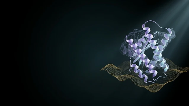
 AI Structure Prediction
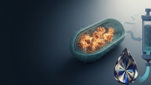
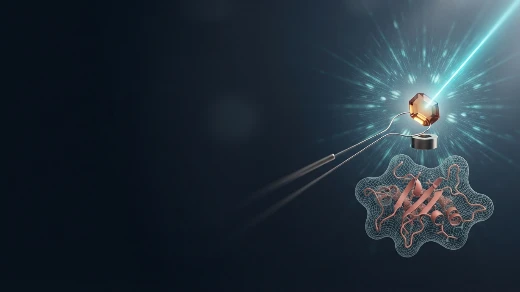
 Multi-Modal Structure Determination
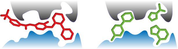
 Integrated CADD
Structure-based drug design lives or dies on the quality of your target structure and the speed at which you turn coordinates into optimized leads. Our platform unifies AI-driven structure prediction, experimental structure determination, and computational drug design under a single project team — eliminating the vendor fragmentation that kills SBDD timelines.
Creative Biostructure at a Glance
Over a decade of trusted expertise powering biotech, pharma, and research institutions worldwide to advance therapeutic innovation.






Why Partner With Us
Most SBDD programs collapse at the interface between structure and design. A crystallography vendor delivers PDB files; a CRO runs docking; your medicinal chemist sits in between, translating electron density into SAR hypotheses without ever speaking to the person who solved the structure. We built this platform to eliminate those gaps: the same scientists who purify your protein, collect diffraction data, and build your model also run the molecular docking, FEP calculations, and MD validation that turn coordinates into compounds.
Your CapEx is in chemistry and biology. Ours is in structural infrastructure and computational horsepower.
| Stage | What We Deliver | What You Don't Need to Build |
|---|---|---|
| AI Target Assessment | AlphaFold3 druggability scoring; construct design; expression feasibility prediction | Structural biology team |
| Structure Determination | X-ray, Cryo-EM, or NMR — modality selected by target properties, not vendor availability | Synchrotron access, Krios facility, NMR magnet |
| Hit Identification | Virtual screening, fragment screening, and biophysical validation with structural feedback | Screening library, SPR infrastructure |
| Lead Optimization | Docking-guided SAR; FEP affinity ranking; co-crystal structure validation | CADD software licenses, GPU cluster |
| Data Handoff | Series of ligand-bound structures + optimized compound set with binding mode documentation | — |
Production-Ready Deliverables: Every campaign ships with validated target structures, hit-to-lead compound series, ligand-bound complex coordinates, and a structural rationale for patent filings.
- ✓ Milestone-based pricing aligned with your fundraising cycles
- ✓ No vendor coordination overhead — protein production, structure determination, and computational design under a single project manager
Membrane proteins. Large complexes. Transient conformations. "Undruggable" is our starting point.
Proven track record where others fail
GPCRs, ion channels, multi-subunit assemblies, and nucleic acid-protein complexes — targets that crash standard SBDD pipelines due to flexibility, low expression, or lack of diffracting crystals.
Multi-modal structure determination
When crystals refuse to grow, we pivot to Cryo-EM; when targets are too small for EM, we deploy NMR; when experimental structures are intractable, we deploy AlphaFold3-guided homology modeling with experimental validation — all without restarting the project clock.
IP firewall & encrypted data infrastructure
Full audit trails, GLP-ready documentation, client retains 100% ownership of all structures, coordinates, and compound data.
Core Service Modules
Service Module At-a-Glance
| Service | Core Capability | Key Resources & Equipment | Typical Timeline |
|---|---|---|---|
| Recombinant Protein Production | AI-guided construct design; multi-system expression; crystallization-grade purification | All commercial vectors; GST/His/MBP/Flag/SUMO tags; GPCR/kinase/epigenetic inserts; bacterial/yeast/baculovirus/mammalian systems; AKTA FPLC/Avant; LC/MS (TOF) | 2–4 weeks |
| X-ray Crystallography | High-throughput screening; co-crystallization & soaking; automated crystal imaging; home-source & synchrotron collection | X-ray diffractometers; nano-liter crystallization robots; high-throughput liquid handling robots; Rigaku CrystalMation with Minstrel HT UV; synchrotron radiation access | 4–12 weeks |
| Cryo-EM Technology | Negative stain QC; automated vitrification; single-particle analysis; AI-driven 3D reconstruction | TEM (JEOL JEM-2200FS); Vitrobot (FEI); Vacuum Evaporator (JEOL JEE-4B); access to high-quality Cryo-EM facilities | 4–8 weeks |
| CADD & Computational Design | Molecular docking; structure-based virtual screening; MD simulation; binding free energy calculation | High-performance computing cluster; professional software tools and databases | 2–16 weeks |
Recombinant Protein Production
AI-Guided Expression from Construct to Crystal-Grade Material
Key Features:
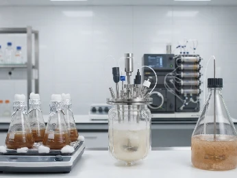
- Multi-System Expression — Bacterial, yeast, baculovirus, and mammalian systems selected by AlphaFold3-guided host prediction; validated inserts for GPCRs, kinases, and epigenetic targets.
- Purification & QC — AKTA FPLC and Avant systems for IMAC, IEX, and SEC; LC/MS (TOF) molecular weight confirmation; NanoDSF/DLS quality gates at every stage.
What We Offer: For virtual biotechs, this eliminates the need to build molecular biology and fermentation infrastructure. For pharma, our GPCR and kinase expression track record de-risks target portfolios from day one.
X-ray Crystallography
High-Throughput Structure Determination from Screening to Coordinates
Key Features:
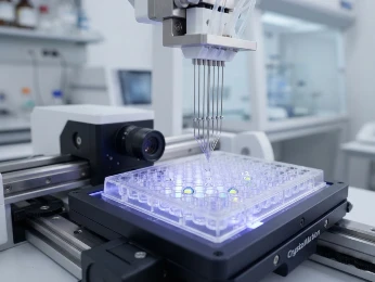
- Automated Screening — Nano-liter crystallization robots and high-throughput liquid handling screen 1,696+ conditions per target; Rigaku CrystalMation with Minstrel HT UV for automated incubation, imaging, and hit scoring.
- Co-Crystallization & Soaking — Sitting-drop and hanging-drop vapor diffusion; ligand soaking with controlled cryoprotectants; flash-cooling at 100 K.
- Data Collection — Home-source X-ray diffractometers for in-house screening; synchrotron radiation facility access for high-resolution datasets.
What We Offer: Our high-throughput crystallography service supports rapid determination of multiple target-ligand crystal structures per month — the throughput essential for iterative lead optimization and SAR validation.
Cryo-EM Technology
Single-Particle Analysis Without Crystallization
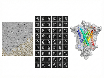
Key Features:
- Sample Prep & Vitrification — Negative stain TEM screening (JEOL JEM-2200FS) for quality assessment; Vitrobot (FEI) automated vitrification; Vacuum Evaporator (JEOL JEE-4B) for grid preparation.
- AI Reconstruction — Access to high-quality Cryo-EM facilities; deep learning particle picking; cryoSPARC 3D classification; cryoDRGN-AI heterogeneity analysis for flexible targets.
What We Offer: For membrane proteins and large complexes that resist crystallization, Cryo-EM bypasses the crystal bottleneck entirely — delivering SBDD-ready coordinates at 2.5–4.0 Å without months lost to crystallization trials.
CADD & Computational Design
From Structure to Optimized Compound — In Silico
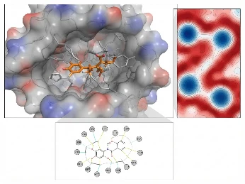
Key Features:
- Molecular Docking & Virtual Screening — Protein-ligand, protein-protein, and covalent docking; AI-enhanced virtual screening against experimental binding sites.
- Molecular Dynamics & Free Energy — All-atom MD and membrane protein MD; FEP/TI and MM/PBSA for lead optimization affinity ranking.
What We Offer: Because our CADD team shares the same lab as our crystallographers, every docking pose is validated against experimental binding modes — not computed in isolation. For hit-to-lead, FEP-guided analog ranking reduces synthesis cycles by 30–50%.
Technology Platform
Integrated SBDD Infrastructure: AI Prediction + Structure Determination + Computational Design, Zero Handoffs
Traditional drug discovery fragments target validation, structure determination, and lead optimization across separate vendors. Our platform unifies all three under one project team, with AI predictions informing experimental design and every structure feeding directly into computational campaigns.
Computational Platform — Dry Lab
| Capability | Details |
|---|---|
| AI Structure Prediction | AlphaFold3, RoseTTAFold, and homology modeling for target assessment, construct boundaries, and druggability scoring |
| Geometric Deep Learning | Graph neural networks and equivariant models for virtual screening, de novo design, and affinity prediction |
| Molecular Docking & Dynamics | Protein-ligand, protein-protein, and covalent docking; all-atom MD and membrane protein MD |
| Free Energy Calculations | FEP/TI and MM/PBSA for lead optimization affinity ranking and selectivity profiling |
| Encrypted Data Infrastructure | Client-isolated project folders; full audit trails; GLP-ready documentation |
Experimental Platform — Wet Lab
| Capability | Details |
|---|---|
| Protein Production | Gene-to-Protein expression in bacterial, yeast, baculovirus, and mammalian systems; isotope labeling for NMR; nanodisc assembly for membrane proteins; AKTA FPLC / Avant FPLC purification; LC/MS (TOF) QC |
| X-ray Crystallography | Rigaku XtaLAB Synergy-R home source; nano-liter crystallization robots; high-throughput liquid handling; Rigaku CrystalMation with Minstrel HT UV imaging; synchrotron partnerships (APS, SSRL, ESRF) |
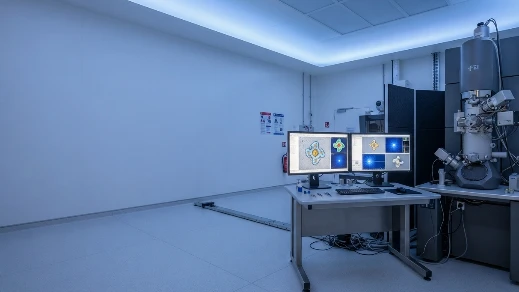
| Cryo-EM | Thermo Fisher Krios G4i (300 kV, Falcon 4i, Selectris X); Glacios 2 (200 kV) for screening; Vitrobot Mark IV vitrification; JEOL JEM-ARM200F for Micro-ED; GPU cluster for real-time 2D classification |
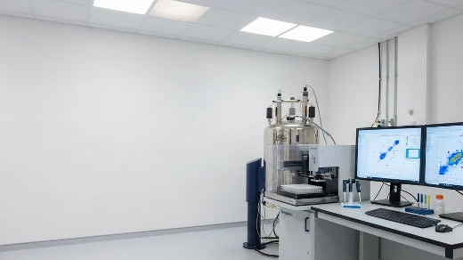
| NMR Spectroscopy | Bruker Avance NEO 800 MHz and 600 MHz spectrometers; QCI cryoprobes (¹H/¹³C/¹⁵N/²H); SampleJet 96-position autosampler; 1.7 mm microprobe for micro-scale samples |
| Biophysical Validation | NanoDSF, DLS, SPR, ITC, BLI for hit validation and QC |
Platform Edge: The ability to assess target druggability with AlphaFold3 on Monday, express and purify the protein on Tuesday, collect diffraction or cryo-EM data by Thursday, and deliver docking-ready coordinates by Friday — all under one project team — compresses traditional 6-month SBDD startup phases into 4-week iterations.
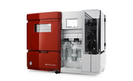
AKTA Avant FPLC
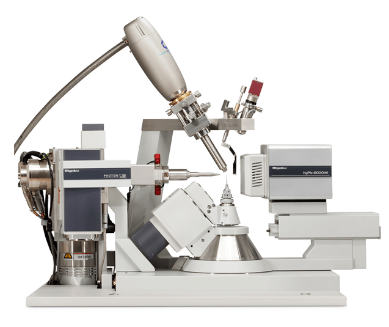
Rigaku XtaLAB Synergy-R
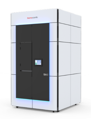
Thermo Fisher Krios G4i
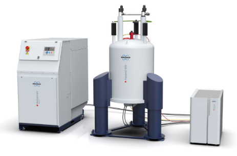
Bruker Avance NEO 800 MHz
Platform specifications are subject to continuous upgrade. Contact our team for instrument availability and project-specific capability assessment.
Closed-Loop Discovery Engine
When AI Prediction Meets Experimental Structure Truth
Static AI models predict single-state structures from sequence. Experimental structure determination reveals the conformational reality that models miss — ligand-induced changes, domain rotations, and ordered water networks. Our platform feeds every experimental structure back into the design cycle.
AI Target Assessment
AlphaFold3 predicts structure, pocket druggability, and construct boundaries; informs expression strategy and crystallization design
→ Feeds into Protein Production
Experimental Structure Determination
X-ray, Cryo-EM, or NMR delivers atomic coordinates; ligand-bound structures reveal binding mode
→ Feeds into CADD
Computational Hit-to-Lead
Docking, FEP, and MD optimize compound series; predicted analogs prioritized by synthesis feasibility
→ Feeds into Validation
Structural Validation
Co-crystal or cryo-EM structures of lead compounds validate predicted binding modes; experimental data refine AI models for next campaign
→ Feeds back into AI
Industrial Value:
For Biotechs
Your first campaign's structural data trains the AI models for your second target. Experimental validation from Phase 0 becomes training data for Phase 1 — a compounding learning partnership.
For Pharma
Every structure and compound is linked to an experimental outcome with project ID, timestamp, and model version — fully audit-ready for regulatory submissions and internal portfolio reviews.
Project Management & Execution
Project Workflow
A standardized, milestone-driven execution system. From sequence intake to lead-optimized compounds.


01 Target Assessment
- AlphaFold3 druggability scoring; construct design; expression feasibility prediction
Deliverable: Target assessment report with construct strategy
02 Gene-to-Structure
Deliverable: PDB/EMDB coordinates; validation statistics
03 Hit Identification
- AI-enhanced virtual screening; fragment screening; docking prioritization
Deliverable: Ranked hit list with structural rationale
04 Hit Validation
- Biophysical validation (SPR, ITC); co-crystal or Cryo-EM binding mode confirmation
Deliverable: Validated hits with KD / binding mode data
05 Lead Optimization
Deliverable: Lead series with FEP affinity estimates
06 Deliverables & Handoff
- Lead compound series; ligand-bound structures; binding mode report; handoff to ADMET / In Vitro ADME-Tox
Deliverable: Final report + patent-supporting structural data + transition plan
Sample Requirements
| Sample Type | Specification |
|---|---|
| Target Sequence | FASTA format; UniProt ID; domain boundaries; post-translational modification requirements; species of origin |
| Protein Material | >95% purity; >0.5 mg/mL; NanoDSF Tm >45°C; DLS PDI <1.2 (if customer-provided) |
| Compound Library | 10 mM DMSO stock; aqueous solubility >50 µM; purity >95%; structural files (SDF/MOL2) |
| Prior Structural Data | Any known homologs, previous models, or published structures for MR template identification |
| Structural Endpoint | X-ray, Cryo-EM, NMR, or biophysical assay |
Standard Deliverables
- Validated target structure (PDB/EMDB coordinates with full validation statistics)
- Hit compound series with AI-ranked binding mode rationale
- Lead-optimized compound set with FEP-validated affinity estimates and selectivity profiles
- Ligand-bound complex structures for patent and regulatory support
- Structural rationale document for SAR and IP filings
- Direct handoff to ADMET Prediction & Modeling, In Vitro ADME-Tox Profiling, or Lead Preclinical Development
Our technical team responds within 24 hours. All inquiries protected under NDA.
Frequently Asked Questions
Case Study
Case #1: Cryo-EM Single-Particle Analysis for Protein Structure
When crystallization fails for large complexes or membrane proteins, cryo-EM provides the only path to atomic coordinates. We executed a complete single-particle analysis workflow — from vitrified grid preparation and Cryo-TEM data collection through AI-driven 2D classification, 3D conformational sorting, and model building — delivering a refined 3D reconstruction at 9.7 Å resolution. The pipeline resolved discrete conformational states from a heterogeneous particle population, demonstrating our platform's ability to turn intractable targets into SBDD-ready structural models without crystal optimization delays.
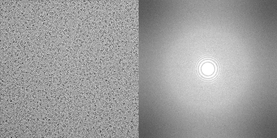
Figure 1. Cryo-TEM data collection: raw micrograph of vitrified protein particles used for single-particle analysis.
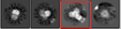
Figure 2. 2D classification: representative class averages revealing particle orientations and overall structural integrity.
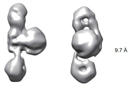
Figure 3. Model building and refinement: final 3D reconstruction resolved to 9.7 Å, displaying the overall molecular architecture.
Case #2: X-ray Crystallography for Multi-Subunit Protein Complex
For targets that crystallize readily, X-ray crystallography remains the gold standard for atomic-resolution ligand-bound structures. We screened crystallization conditions, identified diffracting crystals, and collected synchrotron-grade data to 2.80 Å resolution — processing the diffraction pattern into a high-quality electron density map and building a complete multi-subunit complex structure. The resulting coordinates feed directly into our Molecular Docking and Virtual Screening pipelines, providing the structural foundation for hit identification and lead optimization.
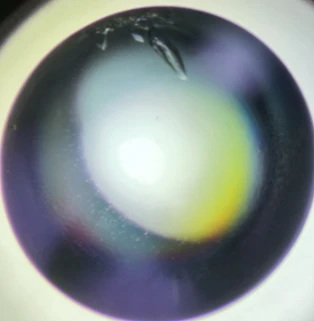
Figure 1. Crystal image: high-quality protein crystal selected for X-ray diffraction data collection.
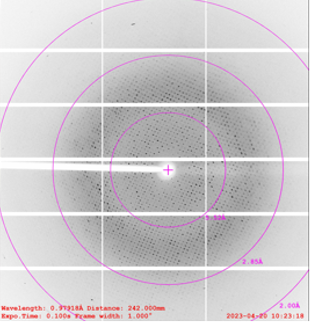
Figure 2. Crystal diffraction: diffraction pattern collected at 2.80 Å resolution, confirming data quality for structure solution.
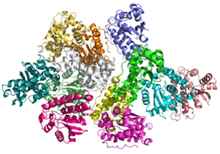
Figure 3. Protein structure: three-dimensional structure of the multi-subunit complex determined at 2.80 Å, with subunits shown in rainbow colors.
Need an integrated SBDD platform to accelerate your drug discovery program? Our team can design a customized structural biology and computational chemistry pipeline tailored to your target class, portfolio timeline, and regulatory milestones. Contact our scientific team today.