Structure Refinement & Quality Assessment
 Structure Refinement
  Low-Resolution Rescue
  SBDD-Ready Validation
 Draft models with poor geometry actively mislead drug design. We rescue them into drug-grade coordinates through MD equilibration, crystallographic refinement, and AI quality scoring.
Why Structure Refinement Is the Critical QA Gate
A structural model with poor geometry is worse than no model — it actively misleads medicinal chemistry. A flipped peptide bond sends docking algorithms chasing phantom pockets; a misplaced loop buries the real binding site. Our platform treats refinement as a drug discovery prerequisite: we combine MD equilibration to resolve physical clashes, crystallographic refinement to improve map-to-model fit, and AI quality scoring to flag regions requiring experimental attention. The result is a model you can trust for virtual screening, lead optimization, and regulatory documentation.
What Sets the Platform Apart
MD + Crystallography + AI
Standard refinement uses either physics or statistics. We integrate both: MD resolves physical clashes, crystallographic refinement optimizes map-to-model fit, and AI scores flag remaining issues.
Drug-Grade Geometry
We optimize for binding pocket shape and side-chain rotamers that matter for docking and FEP — not just global statistics that look good on paper but fail in SBDD.
Experimental Rescue
When refinement reveals weak regions, our structural biology pipeline advances the target to experimental rescue rather than computational patching.
Technology Suite
MD Equilibration & Clash Resolution
Physics-Based Geometry Optimization

Key Features:
- All-atom MD relaxation resolves steric clashes and optimizes hydrogen bonding, improving MolProbity scores by 20–40%.
- Restrained equilibration preserves high-confidence regions while allowing weak loops to relax.
- Explicit solvent MD optimizes water positions and ion coordination critical for docking accuracy.
Ideal For: Homology models with clash issues; Cryo-EM models with flexible regions; preliminary crystal structures with poor geometry.
What We Offer:
Virtual biotechs turn downloaded PDB files into reliable drug design templates. Pharma teams rescue legacy structures that have blocked programs for months.
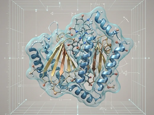
Crystallographic Refinement
Map-to-Model Optimization
Key Features:
- Maximum-likelihood refinement with TLS, B-factor optimization, and anisotropic correction.
- Covalent and non-covalent ligand restraints with dictionary generation for chemically realistic ligand geometry.
- Automated water placement and validation critical for docking and FEP accuracy.
Ideal For: Crystal structures requiring resolution extension; ligand-bound complexes; structures for PDB deposition and publication.
What We Offer:
Our crystallographers combine automated pipelines with manual inspection to optimize R-factors, geometry, and map quality. For lead optimization, we ensure ligand-bound structures are refined to the highest standards.
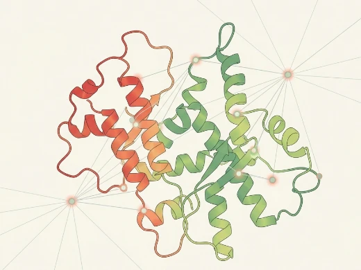
AI Quality Assessment & Experimental Rescue
From Scores to Real Data
Key Features:
- ML classifiers predict model quality from geometric descriptors, identifying high-error regions.
- EMRinger scoring validates Cryo-EM models against map density.
- Construct redesign and co-crystal soaking target weak regions for experimental stabilization.
Ideal For: Model quality assessment before SBDD; prioritization of regions for experimental rescue; regulatory documentation.
What We Offer:
You receive per-residue confidence scores and pocket-specific reliability maps. When computational refinement reaches its limits, we solve it experimentally through our integrated structural biology pipeline.
Platform Instrumentation
Core Instruments
| Instrument | Capability |
|---|---|
| NVIDIA DGX A100 | MD equilibration, AI quality scoring, ensemble analysis |
| NVIDIA RTX A6000 Cluster | Real-time refinement visualization and quality assessment |
| GROMACS/AMBER HPC | Microsecond-scale MD; explicit solvent equilibration |
| Phenix/Refmac Suite | Crystallographic refinement with maximum-likelihood optimization |
| Bruker AVANCE NEO 600 MHz | NMR validation of refined loop conformations |
| Rigaku XtaLAB Synergy | X-ray data collection for experimental rescue |
| Thermo Fisher Krios G4 | Cryo-EM SPA for large complex and membrane protein rescue |
Standardized Workflow
Project Workflow
A milestone-driven execution system from draft model to drug-grade coordinates.


01 Model Assessment
1. Initial model quality assessment with MolProbity and EMRinger
2. Geometry statistics and pocket-specific reliability mapping
Deliverable: Quality report + problem region identification
02 MD Equilibration
1. All-atom MD equilibration with explicit solvent
2. Restrained MD preserving high-confidence regions
Deliverable: MD-refined model + clash resolution report
03 Crystallographic Refinement
1. Phenix/Refmac crystallographic refinement
2. TLS and B-factor optimization
Deliverable: Refined model + R-factor statistics
04 AI Quality Scoring
1. AI quality scoring with per-residue confidence
2. Drug-grade metrics: pocket reliability, docking suitability
Deliverable: Quality report + pocket reliability map
05 Experimental Rescue
1. Construct redesign for experimental rescue (if needed)
2. Co-crystal soaking or Cryo-EM data collection
Deliverable: Drug-grade coordinates + final report
Sample Requirements
- Initial Model: PDB or mmCIF format; homology model, crystal structure, or Cryo-EM model
- Experimental Data: Structure factors (MTZ), EM maps (MRC), or raw data (if available)
- Ligand Information: Known binders, cofactors, or allosteric modulators (for ligand refinement)
- Project Background: Target class, intended use (docking, FEP, publication, regulatory), known quality issues
Standard Deliverables
- Refined structural model with improved geometry statistics (PDB)
- MD equilibration trajectory and analysis report
- Crystallographic refinement statistics (if applicable)
- AI quality assessment report with per-residue confidence scores
- Pocket reliability map for docking and virtual screening suitability
- Experimental rescue data (if applicable): X-ray or Cryo-EM coordinates
- Final technical report with quality metrics and SBDD recommendations
- Electronic data package (refined models, trajectories, analysis scripts)
Frequently Asked Questions
Case Study
Case Study: Multi-domain O-GlcNAcase Structures Reveal Allosteric Regulatory Mechanisms
Goal: Resolve full-length OGA architecture and map how its pHAT domain allosterically regulates catalytic activity through long-range conformational coupling.
Key Data:
- Integrated pipeline: 1.8 Å crystal structure + 3.3–4.0 Å cryo-EM conformations + SAXS validation, with AlphaFold3 models refined via Namdinator and Phenix.
- pHAT architecture: Forms symmetric homodimers with a conserved peptide-binding cleft but no acetyl-CoA binding, classifying it as a pseudo-HAT.
- Four conformations: Human OGA adopts states I–IV where proline-rich linkers tether pHAT domains; pHAT positioning drives arm-region remodeling that reshapes the active site.
- Functional proof: pHAT deletion or linker mutations disrupt O-GlcNAc homeostasis in cellular assays, confirming allosteric regulation.
Why it matters: For drug developers targeting multi-domain or allosteric proteins, this study demonstrates that AlphaFold3 + cryo-EM + MD refinement can resolve flexible regulatory architectures invisible to static crystallography. The discovery that pHAT domains allosterically gate the active site via a dynamic linker-arm mechanism opens a new therapeutic axis—disrupting domain cooperativity for greater specificity.
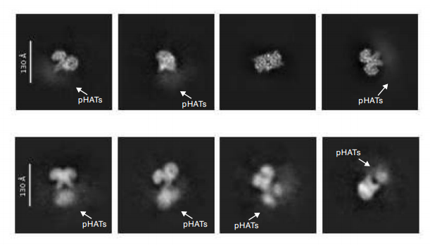
Figure 1. 2D classes reveal ordered catalytic cores with blurred, heterogeneously positioned pHAT domains. (Hansen SB.; et al, 2025)
Reference
Hansen SB, et al. Multi-domain O-GlcNAcase structures reveal allosteric regulatory mechanisms. Nat Commun. 2025 Oct 3;16(1):8828.
Our technical team responds within 24 hours. All inquiries protected under NDA.